|
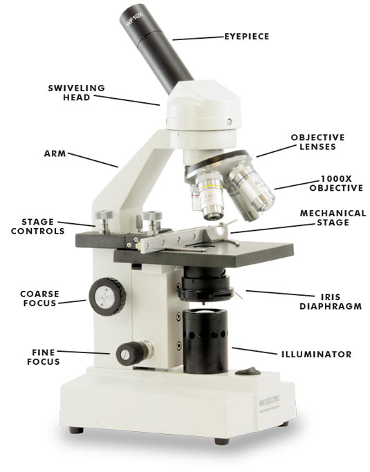
Diagram of a MicroscopeEyepiece - The lens that the microscope operator looks through.
Objective Lenses - Different focus lenses used to see the detail of a specimen, they are the lenses closest to the specimen. Mechanical Stage - A platform where the slide with the specimens are positioned with the clips to hold it in place. Coarse Focus Knob - Used to focus the microscope on low power. Fine Focus Knob - Sharpens the focus on high power. Stage Controls - Moves the stage for the operator to be able to view different parts of the specimen. Swivelling Head - Moves the eyepiece to give the operator better access. Arm - Holds the body tube and can be used for a handle for holding the microscope safely when carrying. Illuminator - The light used to see the specimen clearly, the light shines through the stage. Used instead of a mirror and light. |
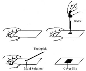
Producing a Wet-MountTo produce a wet-mount firstly you place a few droplets of water onto a slide, secondly place the specimen you want to view onto the slide with the water. Lastly place the cover slip over the specimen by slowly placing it done from one side as not to get any water bubbles left within the cover slip.
|
|
|
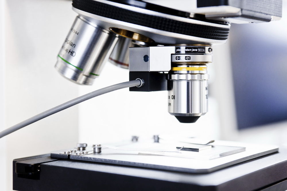
Bringing a Specimen into FocusTo bring a specimen into focus you need to start the stage at the bottom and slowly bring the stage up with the coarse or fine adjustment knobs, being careful when the slide gets close to the objective lens not to crash them together and possibly break something.
You can then move the stage controls to get the best view possible of the specimen or the area you want to look at. |
Drawing a SpecimenWhen drawing a specimen you only need to draw a few of the cells that are being seen through the eyepiece of the microscope being used.
You also need to include the name of the specimen, the date when viewed, a description of the specimen and the magnification level of the objective lens. |
|
